
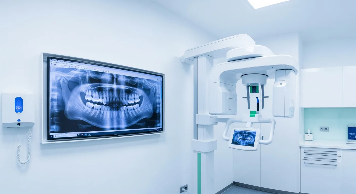
3D X-ray with digital volume tomography
Before a treatment, all risks should be identified as far as possible – volume tomography makes this visible in three dimensions.
Digital diagnostics as the basis of every plan
We use innovative diagnostic techniques: imaging diagnostics are carried out using low-radiation digital X-ray or three-dimensional digital volume tomography (CBCT). Thanks to spatial views, this technique lets dentistry look into new dimensions.
What the third dimension makes visible
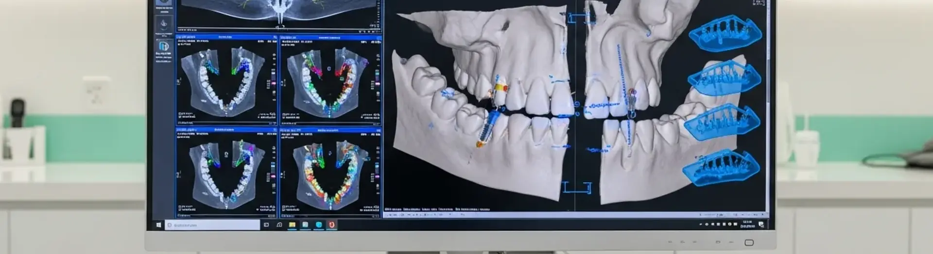
When planning your dentures, the 3D X-ray device allows a considerably better assessment of the surrounding bone. Even before a procedure we obtain precise information about the bone situation and bone quality as well as about the neighbouring anatomical structures. The course of the nerve and the floor of the maxillary sinus can be depicted exactly.
Digital volume tomography enables a spatial, distortion-free and metrically exact representation – a basis for planning that cannot be achieved with two-dimensional images.
Where 3D diagnostics help us further
Implant planning
The position, length and diameter of the implant are determined before the procedure.
Unclear symptoms
Causes can be located and analysed better than in a 2D image.
Root canal system
The three-dimensional course of the canals becomes visible – important for the prognosis of a tooth.
Minimising risks
The position of nerves and the maxillary sinus is known before every surgical procedure.
Added safety with a low radiation dose
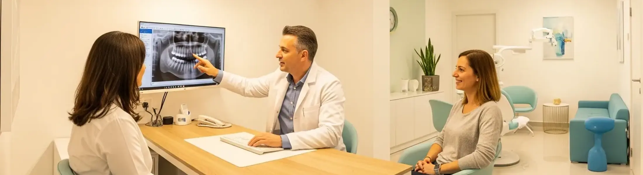
We identify inflammation at the root tip and particular features of the root canal system before treatment begins and can then treat them specifically under the surgical microscope. This adds a clear measure of safety.
The radiation exposure is considerably lower than that of a comparable CT scan. At the same time we gain spatial insights into regions and viewing angles that would not be possible with conventional 2D X-ray technology. Naturally we only take a volume tomography scan when it is medically justified.
The most important points at a glance
- We work with low-radiation digital X-ray and with three-dimensional volume tomography (CBCT).
- Volume tomography shows the available bone, the course of the nerve and the maxillary sinus spatially and without distortion.
- It is the basis for implant planning, root treatments and the clarification of unclear symptoms.
- The radiation exposure is considerably lower than that of a comparable computed tomography scan.